Elizabeth Peters, MD, likes the medical detective work that comes with practicing radiology. Since joining the Emerson medical staff 20 years ago, she has seen the department grow and the technology steadily advance. Dr. Peters, who is married to Richard Kaiser, MD, a psychiatrist on Emerson’s courtesy staff, appreciates the diversity of practice at Emerson and enjoys the regular, productive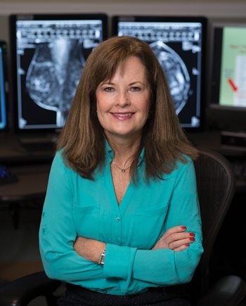 collaboration with her colleagues.
Describe your radiology practice.
I often spend two days a week on mammography at Emerson’s Concord or Westford sites. The rest of the time, I read CT, x-rays, fluoroscopic studies and ultrasound. When I am on call weekends and evenings, I am responsible for reading most of Emerson’s radiology exams. I really enjoy the variety. That’s one of the benefits to working at Emerson: you don’t need to have one focus.
Also, we see different patient populations of all ages. I enjoy the contact with patients and with other staff, including our esteemed technologists and support staff. Our team of radiologists works well together and often collaborate. When we have complicated cases, we research them to make the correct diagnosis and proper recommendations.
What is the perception of how radiologists spend their time?
People think we sit in a dark room all day. That is partly true, but I’m often on the phone with other clinicians. They may have a concern they want me to know about. I appreciate getting additional information on the patient; in fact, I may need more detail. The requisition might say “abdominal pain,” but when I speak with the physician, there’s more to the story.
Referring providers are always welcome to review images with us, either by phone or in person. If I diagnose an abnormality, the physician and I may need to discuss it, and sometimes I suggest alternative imaging. For example, in patients who have a lot of headaches, the physician might order a CT scan with contrast. I’ll call them to suggest the imaging be performed without contrast or to get an MRI instead.
Is the controversy over about when screening mammograms should begin and at what frequency?
Today it seems that most physicians have their patients come in for an annual mammogram beginning at age 40. We see the value of screening mammography all the time, because we see fewer cases of advanced breast cancer than in the past. When we detect particularly aggressive breast cancer sooner, the patient has a much better outcome. Now with 3D mammography, we see every millimeter of tissue. It has reduced the number of callbacks, which results in less stress for our patients.
Technology has steadily advanced since you began practicing.
Yes. Mammography is a good example. We used analog technology until 2004, when we went to digital. In 2013, we began offering 3D mammography, and we now use breast MRI as a problem-solver in certain cases. Ultrasound is another good modality for solving problems in breast imaging.
We use ultrasound in the ER and for pediatrics — say, for an abnormal appendix — in order to avoid a CT. We limit radiation exposure in children and adults as much as possible. CT scans have less radiation exposure than before and have advanced in resolution. We perform sagittal, coronal and thin-section CT. All of these advances have translated directly into better care for our patients.
What attracted you to the field?
When we did our rotations in medical school, I was most interested in the problem-solving, visual aspect of radiology. I had considered primary care, pediatrics or dermatology as well. When we rounded on surgical patients, I wanted to see the CT scans or look at the ultrasounds or other studies. Radiology has become increasingly multi-modality. We use a lot of advanced technology to get the right diagnoses. I feel like a medical detective who looks for findings and tries to determine why the patient is having a problem.

